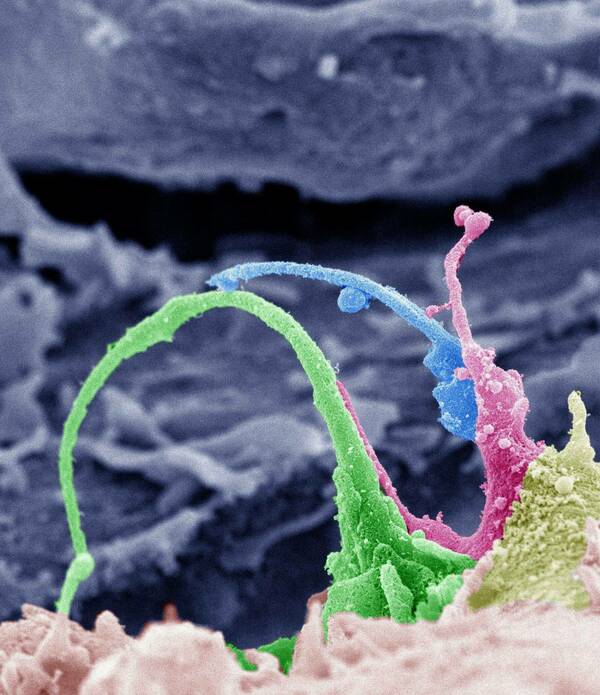
As a recent winner of the Federation of American Societies of Experimental Biology (FASEB) BioArt competition, Jing Yang’s image has been selected for display in FASEB’s BioArt exhibit at the Science Museum of Virginia this summer. The image is a pseudo-colored scanning electron micrograph of surface projections on an ovarian cancer cell obtained in the Electron Microscopy core of the Notre Dame Integrated Imaging Facility. Congratulations Jing!
Image legend: This scanning electron micrograph depicts adhesive structures on the surface of ovarian cancer cells. Early events in ovarian cancer metastasis are regulated by a controlled interaction between cell surface adhesion molecules and proteolytic enzymes that degrade and remove surrounding tissues. Researchers are using images like this one to study the mechanisms by which cancer cells adhere, invade, and metastasize, with the goal of developing novel strategies for early clinical detection and blocking ovarian cancer metastasis.