Breast cancer is the second leading cause of cancer death in women, following lung cancer. Since 1989 the mortality rate of breast cancer has declined due to earlier detection through increased awareness and screening, but a mammogram or CT scan is only as good as the technology used to produce it.
Lisa Cole, a bioengineering PhD candidate, is working at Notre Dame to increase the accuracy of diagnostic tools used to detect microcalcifications during breast cancer screenings. Microcalcifications are small calcium deposits that show up as white specks on mammograms. They can be found in areas of breast tissue where cells are being replaced more quickly than normal, and small clusters can be a sign of pre-cancerous changes.
“My research is focused on investigating the use of a targeted nanoparticle-based X-ray contrast agent to enable contrast-enhanced imaging of breast microcalcifications,” Cole said.
Cole hopes to create a technology that will help radiologists to properly detect a microcalcification and determine the need for a biopsy.
Cole works alongside Dr. Ryan Roeder, Associate Professor in the Department of Aerospace and Mechanical Engineering, and Dr. Tracy Vargo-Gogola, Associate Professor in the Department of Biochemistry & Molecular Biology at the Indiana University School of Medicine, in her research as a part of the Walther Engineering Novel Solutions to Cancer’s Challenges at the Inter-Disciplinary Interface (ENSCCII) Training Project. The ENSCCII Training Project is a fellowship made possible by the generous support of the Walther Cancer Foundation. The program is designed for predoctoral and postdoctoral researchers working on projects that combine cancer research and engineering.
“An engineering background can contribute to addressing cancer by helping to invent novel tools for the improved detection, diagnosis and treatment of cancer. It is going to be the collaborations between multiple disciplines that truly make a difference in addressing cancer, and that is why the ENSCCII training project is such a great opportunity,” Cole said.
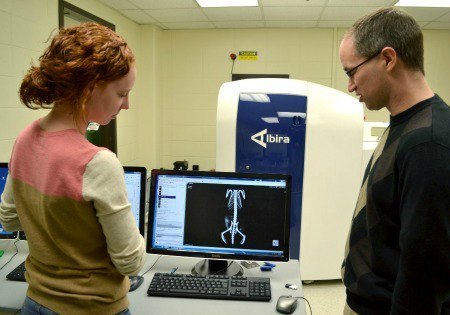
With Dr. Vargo-Gogola and Dr. Roeder as her co-advisers, Cole has been able to utilize several different technologies in her research. For example, Dr. Vargo-Gogola’s lab has extensive experience in animal models. Cole performs different animal experiments, including surgeries and live-animal imaging. In Dr. Roeder’s lab, she uses different instruments to measure the size, surface chemistry and stability of the nanoparticles. This allows her to have great control and knowledge of what is being synthesized.
Dr. Roeder’s lab has previously developed a bisphosphonate-functionalized gold nanoparticle, or BP-Au NP, that can specifically bind to hydroxyapatite mineral. This mineral is the main component of microcalcifications.
“My research has specifically been focused on investigating the ability of BP-Au NPs to enable contrast-enhanced imaging of microcalcifications in in vitro, ex vivo, and two in vivo animals models. The most promising result, so far, is that we can specifically target microcalcifications in vivo and deliver a sufficient mass concentration of BP-Au NPs for contrast-enhanced detection in animal models,” Cole said.
Cole is currently exploring clinical research as a future career path, and she will be spending two weeks at the NIH this summer learning about the role of a PhD in clinical research as well as getting the opportunity to meet principal investigators at the NIH for potential post-doc positions.