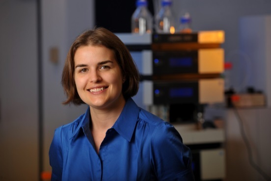
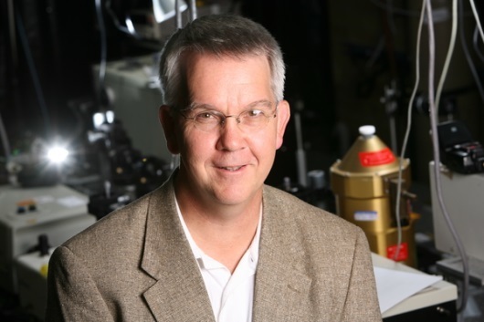
By creating a novel imaging technique, Professor Amanda Hummon, a colorectal cancer specialist, and Professor Paul Bohn, an imaging expert, are looking inside tumors with greater clarity than ever before.
By Michael Rodio | Aug. 8, 2013
One of the most vexing challenges in the fight against cancer is the disease’s adaptability: Even if cancerous cells are quickly detected and treated, they can develop new ways to evade treatment.
To make matters worse, researchers have a hard time “seeing” inside tumors, which are jumbled masses of different kinds of cells. This makes it difficult to test how tumors respond to new drugs designed to overcome that resistance. For doctors, it’s like trying to hit a hidden, three-dimensional target—while the target is moving.
So the Harper Cancer Research Institute created an interdisciplinary team to solve the problem: Amanda Hummon, the Walther Cancer Assistant Professor in the Department of Chemistry and Biochemistry and an expert in colorectal cancer, partnered with Paul Bohn, the Arthur J. Schmitt Professor of Chemical and Biomolecular Engineering and an imaging specialist who directs Notre Dame’s Advanced Diagnostics & Therapeutics Initiative.
Hummon and Bohn pioneered a new imaging tool by combining two forms of 3D imaging: Imaging mass spectrometry (IMS), which shows diagnostically useful proteins in three dimensions, and confocal Raman (CR) microscopy, which displays how specific cell components react to anticancer drugs on a molecular level.
In combination, this new multimodal cancer imaging developed at the University of Notre Dame has potential to revolutionize the way scientists look at tumors in the laboratory—and, with more research funding, it could mean better treatments for cancer patients around the world

